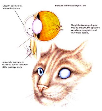
Glaucoma is defined as an increase in intraocular pressure (pressure within the eye). Elevated intraocular pressure (IOP) (greater than 25 mm Hg in the dog and 30 mm Hg in the cat) can lead to pain and irreversible loss of vision. Glaucoma is one of the leading causes of blindness in animals and people.
Symptoms
- Red eye
The vessels on the sclera (the white of the eye) become enlarged, causing the eye to appear red and inflamed.
- Clouding of the cornea ("Corneal edema")
The increased pressure forces fluid forward into the cornea. This disrupts the uniform arrangement of the protein fibers of the cornea, causing them to become edematous or swollen, resulting in a cloudy, bluish-gray color. This is often referred to as a "steamy cornea," similar to the appearance of a bathroom mirror after a hot shower.
- Dilated pupil ("Mydriasis")
Dilation of the pupil, with absence of a response to light stimulation.
- Loss of vision
Vision can be significantly decreased or lost due to the increased pressure. The retina and the optic nerve, which transmits information from the retina to the brain, are damaged by increased pressure. The longer the pressure is elevated, the more extensive and permanent the vision loss may be. Evidence has shown that increased pressure may initiate a chain of events (cellular apoptosis) that persists even after the pressure is relieved, resulting in fiber death within the retina and optic nerve.
- Lens dislocation - ("Lens subluxation or luxation")
The lens of the eye is held in place by fibrous strands or ligaments (lens zonules). The zonules are not very pliable and cannot stretch. With an increase in pressure, they can break, resulting in the shifting of the lens from its normal position.
- Intraocular inflammation - ("Uveitis")
The increased intraocular pressure and subsequent changes within the eye may cause intraocular inflammation (uveitis) to also develop.
- Pain
Elevation in intraocular pressure, especially when acute, may be associated with significant discomfort. People who experience acute glaucoma compare the discomfort with migraine headaches. Similarly, animals with glaucoma may express discomfort with signs such as keeping the eye closed, pawing or rubbing the eye, lethargy, hiding in dim places, restlessness, decreased appetite or vomiting. However, animals are stoic and often don't exhibit all of these signs, or they may only demonstrate them very subtly. Occasionally, decreased activity is attributed to advancing age or arthritis. It is not until the source of the discomfort is alleviated that the animal acts normally again.
- Enlargement of the globe ("Buphthalmia")
Unlike the lens zonules, the sclera can stretch a bit in response to increased pressure. In chronic glaucoma, this can lead to an increase in globe size (buphthalmia). If the pressure increase is prolonged and sustained, the size of the globe will increase because the elastic fibers of the sclera can stretch in response to the build-up of pressure.
Causes
Glaucoma is essentially a plumbing problem and results from a disturbance in the flow of fluid in the eye. Primary glaucoma is usually inherited. Secondary glaucoma occurs when there are other problems within the eye which have contributed to the elevated pressure, such as uveitis (inflammation) or luxation (shifting from the normal position) of the lens.
The fluid that fills the eye, the aqueous humor, is produced by the ciliary body which is located behind the iris. The fluid flows through the pupil, filling the anterior chamber. It normally flows out from the eye through the iridocorneal drainage angle, located where the iris and cornea meet. The ciliary body and the iridocorneal angle encircle the eye, 360 degrees. Normally, the fluid production and outflow are in a steady state, keeping the intraocular pressure in balance. Glaucoma occurs when there is an obstruction to the outflow of fluid. This causes the fluid to build up, thus increasing the intraocular pressure. As the pressure increases, the symptoms of glaucoma can occur.
Primary glaucoma is usually caused by an inherited abnormality in the development of the drainage angle. Breeds particularly susceptible to inherited primary glaucoma include: several Spaniel breeds, including the American Cocker Spaniel and Brittany Spaniel; Basset Hound; Chow Chow; Chinese Shar Pei; Siberian Husky; Norwegian Elkhound; Miniature Poodle; Beagle; Samoyed.
Inherited lens luxation (dislocations). Several terrier breeds, including the Jack Russell and Wirehaired Fox terrier, and the Chinese Shar Pei have an inherited lens zonule deterioration condition which can lead to luxation of the lens. If the lens luxates forward, through the pupil into the anterior chamber, it disrupts the flow of the aqueous humor, causing a significant, rapid increase in the intraocular pressure
Intraocular inflammation (uveitis) can cause:
• Blockage of the drainage angle by inflammatory cells
• Closure of the drainage angle due to the inflammatory process (common in cats - chronic uveitis also a common cause of lens luxation in the cat)
• Adhesions (synechia) of the pupil margin to the lens causing an obstruction of aqueous humor flow (Iris bombe) or to the cornea leading to obstruction of the drainage angle
Intraocular tumor
Diagnosis
The diagnosis of glaucoma is based on a thorough ocular examination and measurement of the intraocular pressure with a tonometer. Gonioscopy is a diagnostic evaluation of the iridocorneal drainage angle, requiring a special lens, which allows its direct visualization. Gonioscopy may sometimes aid in determining the risk of the development of glaucoma in a normal eye, especially in breeds predisposed to the development of glaucoma.
Treatment
A treatment protocol is formulated based on the classification of glaucoma, underlying causes (lens luxation, uveitis, tumor), and the prognosis for vision. Treatment of glaucoma is aimed at control, as glaucoma is rarely cured! A rapid and accurate diagnosis is essential, as an intraocular pressure elevation sustained for a period of just one day, can result in permanent blindness.
Medical
The goal of medical treatment is to decrease production of fluid and increase its outflow from the eye. This is generally achieved by the use of a combination of medications. Here are several categories of medications that are often used in combination:
- Diuretics
Osmotic diuretics such as intravenous mannitol or oral glycerin are used to rapidly decrease the intraocular pressure in acute glaucoma. This is usually administered on an emergency basis with the pet hospitalized for monitoring.
- Beta Blockers
Beta Blockers, such as Levobunolol, Timolol, Metipranolol and Carteolol Hydrochloride work by decreasing the production of fluid by the ciliary body. There is some evidence that they may also increase outflow. Side effects include the possibility of decreased heart rate. This medication should be used with caution in patients with cardiac or respiratory disorders (asthma).
- Carbonic Anhydrase Inhibitors (CAI)
Carbonic anhydrase inhibitors such a methazolamide and dichlorphenamide, taken orally, and Dorzolamide or Brinzolamide applied topically, suppress aqueous humour production by the ciliary body. Use of oral carbonic anhydrase inhibitors may cause panting (due to metabolic acidosis) and cause a decrease in appetite. Vomiting and/or diarrhea can also be seen.
- Beta Blocker / CAI combination
Dorzolamide hydrochloride - Timolol maleate ophthalmic solution combines the benefits of both drugs into the convenience of one solution. Their combined effect lowers intraocular pressure greater than either component administered alone.
- Prostaglandins
Prostaglandins such as Latanaprost ophthalmic solution decrease intraocular pressure by decreasing the pupil size and increasing uveoscleral outflow - a secondary outflow pathway for the aqueous humor.
- Corticosteroids and Non-steroidal Anti-inflammatories (NSAID)
Corticosteroid preparations, either topically or orally, and / or oral NSAID may be prescribed to treat intraocular inflammation.
- Miotics
Cholinergic agents such as Pilocarpine and cholinesterase inhibitors such as Phospholine Iodide may be used to constrict the pupil. These drugs produce miosis (constriction of the pupil) through contraction of the sphincter muscles, causing increased tension on the drainage angle, therefore facilitating aqueous outflow. The use of these drugs often causes ocular irritation and may potentiate uveitis. Miotics may be used in combination with other glaucoma medications.
- Sympathomimetics
Sympathomimetics such as Epinephrine or Dipivefrin are also used in the treatment of glaucoma. They have been documented to enhance aqueous outflow while decreasing aqueous production.
Surgery
Glaucoma is generally considered a surgical disease. Successful management of glaucoma generally depends on the use of medications in conjunction with surgery.
- Diode Laser Photocoagulation
In this procedure, pulses of diode laser energy are applied, circumferentially around the eye, via a probe placed on the sclera above the ciliary body. The laser creates local thermal destruction of the ciliary body. By destroying focal points of the ciliary body, aqueous humor production is reduced - the goal being to restore intraocular pressure equilibrium.
- Cylocryothermy
In this procedure, a glaucoma cryo (freezing) probe is placed over the ciliary body, circumferentially around the globe. This freezing leads to destruction of the ciliary body thus reducing aqueous humour production.
- Gonioimplant
Implantation of an anterior chamber shunt or valve to facilitate outflow of aqueous humor from the eye. The drainage is usually onto the sclera of the eye, under a conjunctival pocket. A small tube leads from the device, through a scleral tunnel, into the anterior chamber. If the intraocular pressure rises above a certain threshold, the excess aqueous is siphoned off via this tube to a filtration bleb. The main cause of failure of these implants is the extensive scarring that can occur around the implant causing the filtration bleb to seal. Gonioimplantation is often performed in conjunction with laser photocoagulation.
Treatments for Non-Visual Eyes
- Chemical Ablation
Intravitreal injection of Gentamicin (or other medications) is toxic to the ciliary body, and leads to its destruction. The procedure often requires mild sedation and aspiration of some fluid from the eye prior to the injection. As the ciliary body dies, the production of aqueous humor decreases, resulting in a decrease in intraocular pressure. The soft globe often atrophies (phthisis bulbi), occasionally shrinking significantly in size. This procedure can be used in end-stage, buphthalmic and blind eyes. As the Gentamicin is also toxic to the retina, it should not be used to treat eyes with a potential to save vision. The Gentamicin injection often causes marked intraocular inflammation, which may become chronic, causing intermittent intraocular hemorrhage.
- Intraocular Prosthesis
In this procedure, the contents of the globe are surgically removed (evisceration) and replaced with a solid silicone sphere to maintain the normal size and shape of the eye. The cosmetic results are variable, and the cornea often becomes cloudy in appearance. This surgical procedure leaves the patient with a comfortable globe and is significantly more cosmetic than enucleation.
- Enucleation
Surgical removal of the eye, with the eyelids permanently sutured closed. A blind, glaucomatous eye suspected of having an intraocular tumor is usually enucleated (vs. having a prosthesis place).
Prognosis
The prognosis for a patient with glaucoma is highly dependent on the early detection and treatment. Due to the nature of the disease many animals eventually lose vision in the affected eye despite treatment. Once the battle for vision in an eye is lost, the goal becomes to maintain a cosmetic, comfortable eye, and prophylaxis or protection of the fellow globe. On average, patients with primary, inherited glaucoma will develop glaucoma in the fellow (normal) eye within twelve months of diagnosis. Successfully managing a pet with glaucoma requires long-term vigilant treatment and frequent evaluations.
Illustration reprinted with permission by the copyright owner, Hill’s Pet Nutrition, Inc.