After a diagnosis of cancer is made, the staging of that patient's cancer is helpful for determining whether it has spread (metastasized) to other organs, or whether it is localized to the area of its origin. This information provides the veterinarian a comprehensive picture from which a prognosis and treatment options can be made that are individualized for that patient.
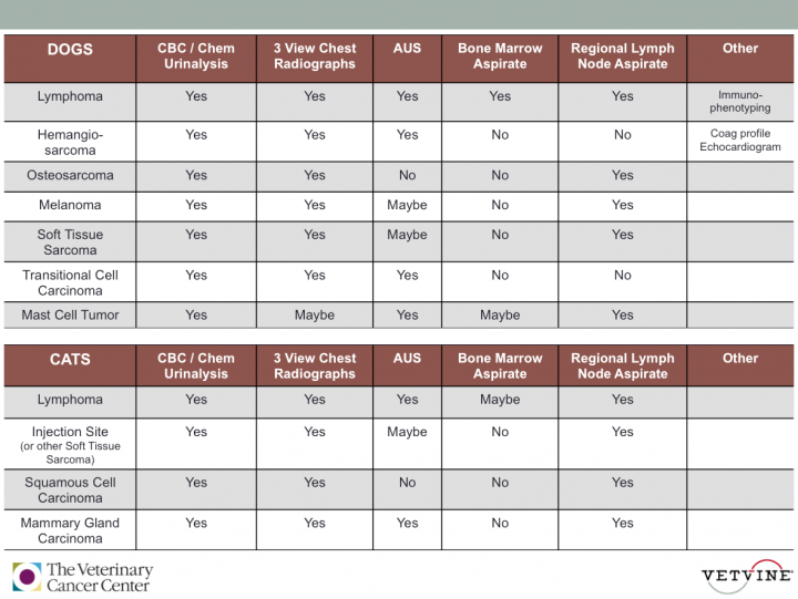
Staging Tests Commonly Performed
- CBC - Complete Blood Count
Prior to chemotherapy treatment for any cancer type, it is necessary to confirm that a patient has enough red blood cells, white blood cells and platelets to safely receive the treatment.
Additionally:
Lymphoma - some patients will have evidence of cancer cells in the bloodstream; Cell counts can be low if there is bone marrow involvement.
Hemangiosarcoma - Some patients may develop changes in the appearance of red blood cells; Some patients can become severely anemic, thus requiring blood transfusions as part of their treatment.
Mast Cell Tumors - Some patients can develop anemia (low red blood cell count); Occasionally there can be decreases or increases in certain cell counts; Mast Cells seen in the circulation can suggest bone marrow involvement or a systemic disease (Mastocytosis).
- Chemistry Panel
Evaluation of internal organ function (especially liver and kidneys) is essential prior to starting any chemotherapy treatment plan. Abnormal test results can be suggestive of abdominal organ involvement.
Additionally:
Lymphoma - This cancer commonly invades the liver, and can also affect the kidneys and gastrointestinal tract.
Hemangiosarcoma - Internal organ dysfunction can be indicative of metastatic disease.
Osteosarcoma - Some patients can have an elevation of ALP (alkaline phosphatase).
- Urinalysis
Urine sample analysis is part of the evaluation of kidney function. This is essential prior to starting any chemotherapy treatment protocol. In addtion, patients with cancer have a diminished immune system, and as a result, can develop urinary tract infections.
Addtionally:
Transitional Cell Carcinoma - Some patients may have kidney failure due to obstruction of the ureter associated with tumor growth; Cancer cells can be seen in the urine in up to 30% of cases; Patients have a higher incidence of urinary tract infection, and bacteria can be seen in the urine.
- Three-view Thoracic Radiographs
Imaging (X-rays) of the chest and lungs can reveal changes consistent with primary tumors or nodules, and/or metastatic cancer. Three views are recommended in cancer patients (instead of the typical two-view study), as a moderate percentage of nodules will not be detected on a one or two-view study. Chest radiographs are usually recommended prior to pursuing aggressive local or systemic cancer therapy - even in patients with cancers known to have low metastatic rates.
Additionally:
Lymphoma - Imaging may reveal a mediastinal mass, enlarged lymph nodes, and/or diffuse, infiltrative disease. Repeating of radiographs during the course of treatment can be helpful for monitoring the remission status and response to treatment.
Hemangiosarcoma - This cancer type has a high metastatic rate and often spreads to the lungs.
Canine Soft Tissue Sarcomas - This tumor type has a low metastatic rate (less than 15% for low grade tumors, and approximately 40% for high grade tumors), however it is important to know if metastatic disease is present prior to recommending aggressive local (radiation) therapy.
Transitional Cell Carcinoma - Although this tumor type has a low metastatic rate (less than 15% in one study), patients have a higher likelihood of spread to the lungs as the disease progresses.
Mast Cell Tumors - Thoracic radiographs are not usually recommended, as it is uncommon for this tumor to spread to the lungs. The exception is if there is a suspicion of spread to the sternal lymph node, if the patient is having problems breathing, or if ruling out pulmonary (lung) disease is warranted.
Melanoma - In dogs, this tumor type has a moderate to high metastatic rate.
Osteosarcoma - This tumor has a high metastatic rate (greater than 90%), and often spreads to the lungs.
Feline Injection Site (or Soft Tissue) Sarcoma - These have a low to moderate metastatic rate (10-25%).
- Abdominal Ultrasound (AUS)
Since some cancers can invade the abdominal organs, a complete abdominal ultrasound (AUS) may be recommended to assess for abdominal involvement. This can help to set the prognosis for some patients.
Additionally:
Lymphoma - Commonly affects the liver, spleen, and abdominal lymph nodes; An AUS can also assess if other organs are affected including the gastrointestinal tract, kidneys, or pancreas.
Hemangiosarcoma - This cancer typically arises from the spleen or liver, and has a high metastatic rate; The AUS can help to assess for the presence of metastatic disease, and/or the presence of abdominal effusion - which can indicate tumor rupture or bleeding.
Mast Cell Tumors - AUS is very important for staging this cancer type as this tumor can spread to the liver, spleen and abdominal lymph nodes; If the liver and/or spleen appear abnormal on the AUS, then an aspirate for cytology is warranted to rule out the presence of metastatic disease; Patients with high grade tumors should always have a fine needle aspirate performed of the liver and spleen (even if the AUS findings appear normal).
Canine Soft Tissue Sarcomas and Feline Injection Site (or other Soft Tissue) Sarcomas - In cases where the tumor develops over the abdomen or hind limbs, an AUS is recommended to evaluate for metastatic disease to internal regional lymph nodes or other abdominal organs.
Transitional Cell Carcinoma - AUS is very important for staging of this cancer as it is common to see involvement of other parts of the urogenital system (ureter, urethra, prostate); It's important to assess the ureters for any obstruction; The regional lymph nodes, liver and spleen can be evaluated for metastasis; The AUS helps to define where in the bladder the mass is located which can impact whether surgery is recommended as part of the treatment plan.
Feline Mammary Gland Carcinoma - Since over 25% of patients will have evidence of lymph node metastasis at the time of diagnosis, an AUS may be recommended to assess the abdominal organs and lymph nodes.
- Bone Marrow Aspirate
A bone marrow aspirate is recommended to check for metastasis or bone marrow involvement in patients with certain cancers. If bone marrow involvement is confirmed, adjustments in treatment protocols may be necessary.
Additionally:
Lymphoma - In patients with an abnormal CBC (Complete Blood Count), a bone marrow aspirate is recommended to check for bone marrow involvement.
Mast Cell Tumors - In dogs with high grade tumors (including aggressive grade II or grade III Mast Cell Tumors), this test may be recommended, as up to 5% of patients will also have bone marrow involvement - this will help determine the best course of treatment for these patients.
- Lymph Node Aspirate
Lymph nodes in the vicinity of a tumor or mass (regional lymph nodes) can serve as site of metastasis (spread of tumor cells). An aspirate of the lymph nodes may be recommended for certain tumor types as part of the staging for that tumor type.
Additionally:
Osteosarcoma - Spread to the regional lymph nodes is not common in dogs (less than 5%), however, if tumor cells are seen in the regional lymph node aspirate, the prognosis is known to be worse compared to dogs without regional lymph node involvement.
Canine Soft Tissue Sarcomas and Feline Injection Site (or other Soft Tissue) Sarcomas - It is important to determine if there is metastasis to regional lymph nodes prior to making treatment recommendations for patients with this tumor type.
Mast Cell Tumors - In dogs, it is common for this tumor to metastasize to regional lymph nodes. Although spread to the lymph nodes does not significantly alter the prognosis, it does help to determine the best treatment plan for that pet; If mast cells are seen on cytology, a lymph node biopsy (or lymph node removal) is often recommended to distinguish an inflammatory response from actual metastasis.
Melanoma - In dogs, it is important to know if regional lymph node spread is present before treamtent recommendations are made for this tumor type.
Feline Squamous Cell Carcinoma - It is important to know if regional lymph node spread is present in order to determine the best course of treatment in affected cats.
- Coagulation Profile
This blood test helps to determine if a patient's blood clots normally. The majority of patients with Hemangiosarcoma show one or more coagulation abnormalities. If clotting abnormalities are found, certain patients may require treatment with various blood products (transfusion) depending on the severity and stability of the patient.
- Immunophenotyping
In patients with Lymphoma, this testing helps to determine the type of lymphoma present (B-cell or T-cell). This is important for guiding treatment options and creating a prognosis for the patient with lymphoma.
- Echocardiogram
This is not a commonly performed staging test except in cases where cardiac (heart) Hemangiosarcoma is suspected. In 9-25% of patients with splenic (spleen) hemangiosarcoma, there can also be heart (right atrium) involvement. This test can help to guide the best course of treatment and assess risks in these patients.
Contributed by: The Veterinary Cancer Center